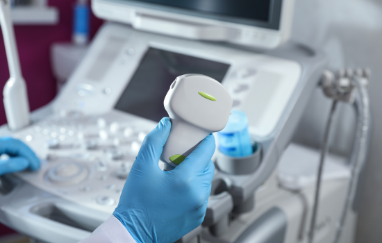
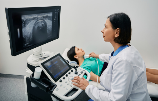
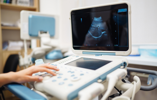
What is 3D Sonography?
3D Sonography (Three-Dimensional Ultrasound) is an advanced imaging technique that provides clear, lifelike images of internal organs, tissues, and developing babies during pregnancy. Unlike traditional 2D ultrasound, which shows flat, grayscale images, 3D sonography captures images from multiple angles to create a three-dimensional view.
Why Choose 3D Sonography?
- High-Resolution Images – Offers detailed visuals of organs and fetal structures.
- Pregnancy Imaging – Allows expecting parents to see their baby’s facial features, limbs, and movements.
- Better Diagnosis – Helps in detecting congenital abnormalities, uterine issues, and organ conditions with improved clarity.
- Safe & Painless – A non-invasive, radiation-free, and completely safe diagnostic procedure.
When is it Recommended?
- During second or third trimester of pregnancy (for fetal development check).
- For gynecological and abdominal assessments.
- To detect structural abnormalities or assist in pre-surgical planning.